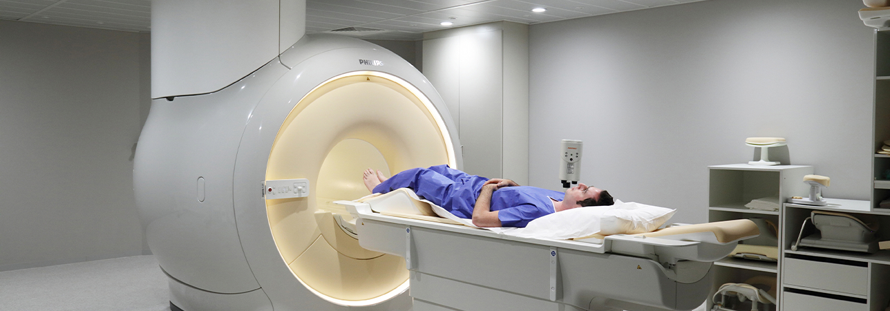
MRI (magnetic resonance imaging) scan in a radiology department.

MRI

Magnetic resonance imaging is a non-invasive technique.

The device consists of a magnet characterised by its power (1, 1.5 or 3 Tesla) and an examination bed which glides down its centre. The examination creates images of the human body thanks to the hydrogen atoms it contains. In a strong magnetic field, all the hydrogen atoms move towards this field. They are then excited by radio frequency waves for a very short period of time.

When the wave emission stops, the atoms return to their steady state and the energy accumulated produces a signal that is recorded and processed as an image. Specific antennas according to the area studied are connected and used to collect the emitted signal. Such signals are mathematically analysed to produce images.

MRI and ultrasound do not use X-rays

According to the technique or sequences, MRI enables the morphology of the organs studied to be brought to light in a detailed way. It demonstrates an increased sensitivity in detecting different pathologies of the soft tissues, making vessels, cartilage and nerve pathways stand out. The acquisition of three-dimensional images allows for their reconstruction on all planes.

These non-radiation examinations use either the properties of magnetic fields (MRI) or the properties of ultrasound. For the intensities used by these two techniques, no specific effects have been observed in humans.

Examination

During your examination, you will be accompanied by a medical radiology technologist (MRT). They will provide you with a private area where you can undress according to the requirements of the examination. In principle, the examination is painless but noisy. You will be offered a headset to listen to music and/or ear plugs.

If preparation is required, do not forget to follow any instructions to ensure that the examination can be carried out correctly and will provide quality results.

A contrast agent injection may be necessary.

You will be offered a temporary gown.

Please do not hesitate to ask any questions and make any requests. We will strive to fulfil them.

The examination will take 40 minutes on average.

Doctors with this specialisation

Clinique de Montchoisi

Dr. med. Sylvain Eminian

Specialisation
Standard radiology, Radiology, MRI, Computed tomography, Ultrasound, Mammography View more
Clinique de Montchoisi

Dr. med. Anne-Lise Hachulla

Specialisation
Radiology, Standard radiology, Mammography, MRI, Computed tomography, Ultrasound View more
Clinique de Montchoisi

Dr. med. Stefano Binaghi

Specialisation
Radiology, Ultrasound, MRI, Mammography, Computed tomography, Interventional radiology, Standard radiology View more
Clinique de Montchoisi

Dr. med. Emmanuèle Cerottini

Specialisation
Radiology, Ultrasound, MRI, Mammography, Standard radiology, Interventional radiology, Computed tomography View more
Clinique de Montchoisi

Dr. med. Yaël Maeder

Specialisation
Radiology, Standard radiology, Mammography, MRI, Computed tomography, Ultrasound View more
Clinique de Montchoisi

Dr. med. Eric Pesquié

Specialisation
Radiology, Ultrasound, MRI, Computed tomography, Mammography, Standard radiology View more

Find a doctor

Our specialists have many years of experience. We guarantee a quick, professionally competent clarification and consultation as well as treatment according to the most modern possibilities.