Treatment of hip injuries and diseases at Swiss Medical Network

Hip surgery

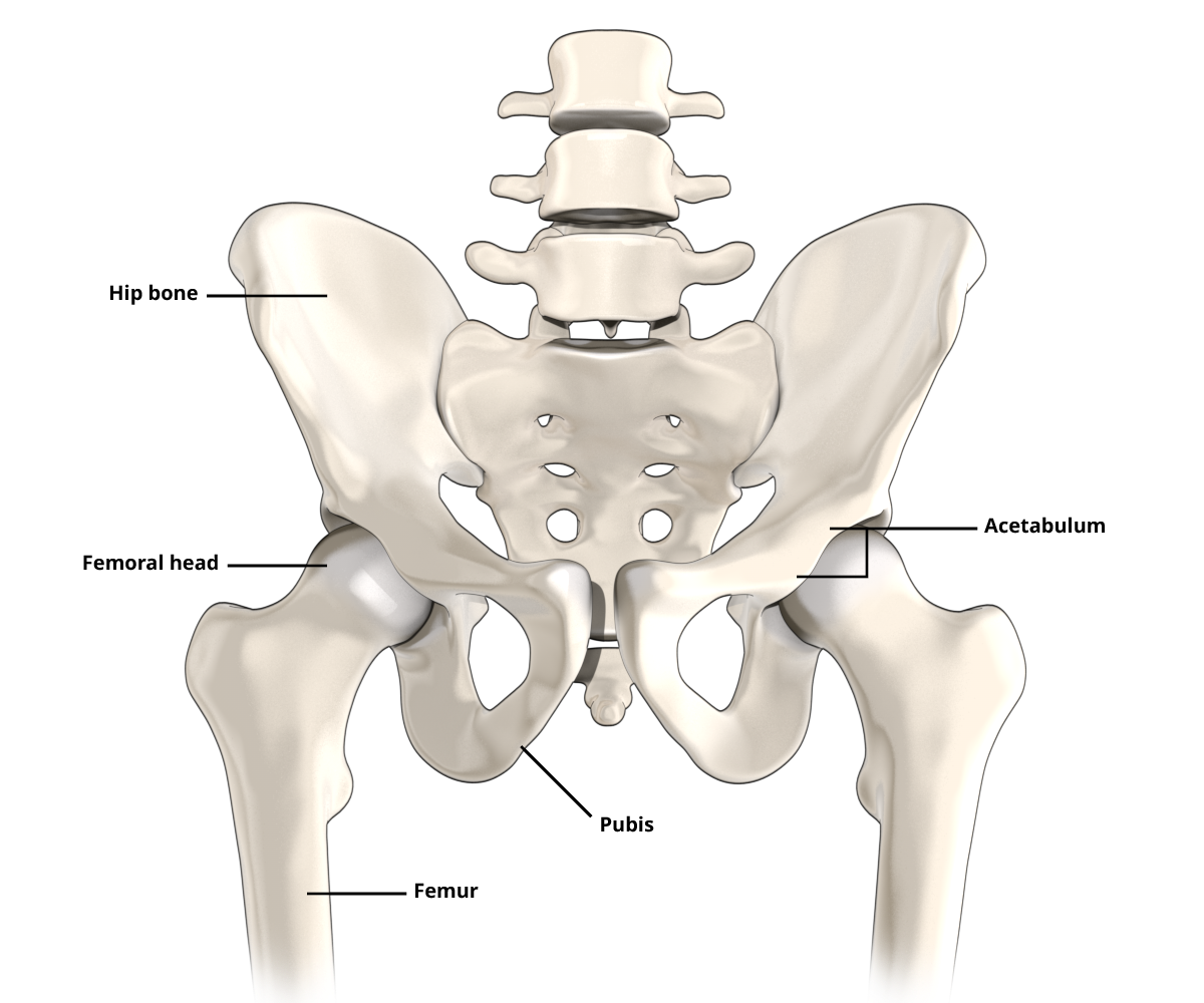
The hip joint is the second largest joint in the human body. The femur and the pelvis come together to form the hip joint. The acetabulum and the femoral head are covered with a resistant cartilage with smooth, sliding surfaces. This allows the hip joint to move with minimal friction. If this cartilage degenerates or becomes injured, low-friction movement can no longer be guaranteed. 

Hip surgery is used to treat all types of hip problem. The treatment methods include both conservative and state-of-the-art surgical measures.

Most common injuries and diseases

Further information on diagnosis, therapy and treatment options

Hip osteoarthritis
Hip impingement
Hip prosthesis

Hip pain | Symptoms

The type of pain and discomfort that occurs in the hip very much depends on the age and physical activity of the person concerned.

Hip pain often coincides with certain movements and is accompanied by restricted mobility. Stinging or electrifying pain can radiate into the thigh. Pain in the outside of the hip and a limping gait are very typical symptoms.

Hip problems can appear suddenly, such as after an accident or over-exertion. They tend to worsen over time and sometimes cause restricted mobility.

When hip pain occurs in older people, it is usually in connection with osteoarthritis, where the cartilage on the hip joint has worn away

Causes

There are various reasons why hip pain might occur. Typical causes are accidents, wear and tear, a congenital deformity, a trapped nerve or an infection.

Here is a selection of the potential causes:

  • broken bone, e.g. femoral neck fracture
  • traumatic hip dislocation (accidental dislocation of the hip joint)
  • arthritis of the hip (inflammation of the hip joint caused by bacteria [septic coxitis, infection], rheumatism [rheumatoid arthritis] or by an inflammatory flare-up [reactive osteoarthritis])
  • hip dysplasia (congenital misalignment of the hip)
  • advanced coxarthrosis (arthritis in the hip joint)
  • hip impingement (painful blockage of the hip joint caused by bone spurs). This is referred to as ‘pincer impingement’ when it occur on the socket and ‘cam impingement’ on the femoral neck. The bony deformity on the acetabulum and/or femoral head impairs the mobility of the hip joint (flexion in particular) and causes pain.)
  • labral tear (tear of the important ring of cartilage that seals the rim of the hip joint)
  • bursitis (inflammation of the bursa), e.g. trochanteric bursitis (bursitis of the greater trochanter)
  • periarthropathia coxae (wear-related disease of the tendon attachment of the stabilising hip muscles on the greater trochanter)
  • coxa saltans, also known as ‘snapping hip’ (caused by a tight tendon [tractus iliotibialis], which gets caught on the greater trochanter when the hip is flexed, causing a jerky, snapping motion.)
  • meralgia paraesthetica (compression of the sensitive lateral femoral cutaneous nerve beneath the inguinal ligament: Those affected suffer from abnormal sensations [paraesthesia] as well as burning pain and sensory disturbances on the front or outer side of the thigh.)
  • Osteonecrosis of the femoral head (death of the bone tissue on the femoral head: This can occur without a recognisable trigger [idiopathic] or as a result of cortisone tablets or injections, alcohol abuse, metabolic disorders or arterial occlusive disease.)
  • perthes disease (necrosis of the femoral head, occurring in childhood)
  • juvenile femoral head release (epiphyseolysis capitis femoris: slipping of the femoral head along the growth plate during puberty.)

Diagnosis

The patient is asked to describe their symptoms in detail so that the surgeon can make an informed assumption of what is causing the pain.

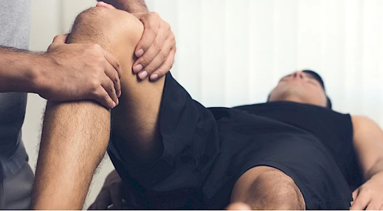
A physical examination is usually carried out to examine possible malpositions of the spine, knee and hip. In addition, there will be a more thorough examination of the patient’s gait pattern, muscle reflexes and the mobility of the hip joint.

In order to confirm the diagnosis, further examinations are then carried out, e.g. blood analysis, X-ray or ultrasound examinations. In special cases, tissue samples are taken. If more precise assessment of certain hip structures is required, CT (computed tomography) or MRT (magnetic resonance imaging) images are taken.

Treatments

Hip pain can be treated conservatively (without surgery) or surgically.

Conservative therapies

  • Ultrasound, shock wave therapy, massage, muscle training, etc.
  • Anti-inflammatory drugs and lubricants
  • Injections of hyaluronic acid or cortisone into or around the hip joint (e.g. for bursitis)
  • Drugs designed to support the cartilage, or appropriate dietary supplements for the joint

Surgical treatment

  • Fixation of fractures with plates, screws or nails
  • Suture of tendon tears
  • Removal of bursa and bone spurs
  • Arthroscopy of the hip joint with suture or removal of the labrum (the cartilage around the hip socket) and removal of bone spurs in the case of hip impingement
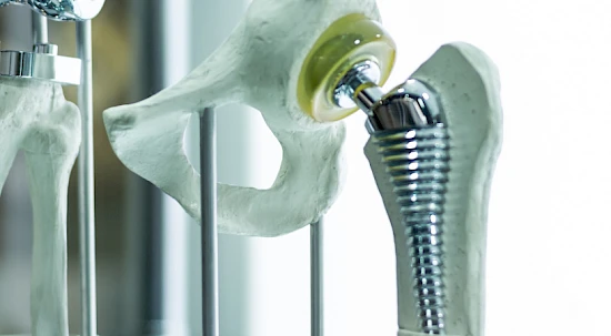
  • Artificial hip replacement: If the osteoarthritis has caused so much wear that the hip joint is destroyed, it can be replaced with an artificial hip joint.
  • During the operation, the surgeon will decide whether to preserve the existing hip or to replace it with an artificial hip joint.

We find a specialist for you

News

La Consulenza Rete Uno - Dr. med. Dario Giunchi

Blog

Artificial joint: How do I decide in favour of (or against) it?

Artificial knee or hip joint: why patient education is key