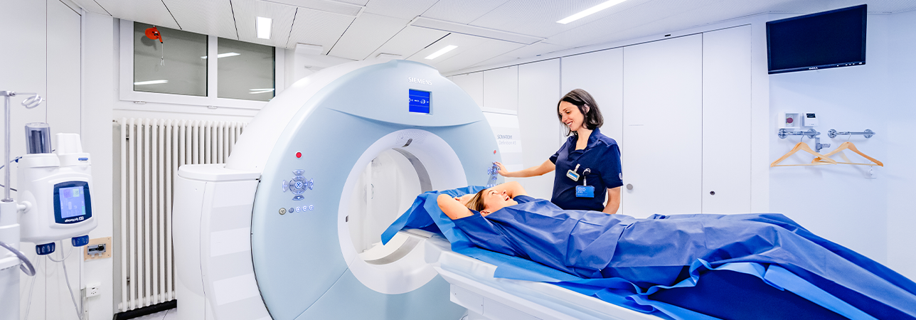
Computed tomography (CT) scan in a radiology department.

Computed tomography

Since 1972, several technological generations of CT scanners have succeeded one another. The state-of-the-art ones enable extensive anatomical regions to be investigated in a very short space of time.

CT scanners are devices that use an X-ray tube rotating around the patient. Sensors scan all or part of the human body and provide data to reconstruct the image numerically.

Area of use

This technique has established itself in all fields of radiology. Its diagnostic contribution is essential, particularly for abdominal pathologies, postoperative monitoring, orthopedic measurements, new virtual endoscopy techniques and vascular pathologies. It is used for virtual colonoscopies in the event of failure of optical colonoscopies, or as an alternative in specific cases. It is also used for coronary computed tomography angiographies, for a precise study of the coronary arteries, within the framework of screening for coronary artery disease, as well as in preoperative assessments and postoperative follow-ups.

CT can also be used for interventions that require precise anatomical identification, such as the treatment of pain via guided lumbar infiltration.

It also enables such identification in order to perform biopsies, drainage and oncological treatments using radio frequency (percutaneous destruction of tumours using heat) or cryotherapy (percutaneous destruction of tumours using cold).

Radiographs and CT scans use X-rays

In the field of irradiation of patients, low doses are used and precautions are taken to keep the area examined to an absolute minimum. For example, on average, a simple image corresponds with the average natural (cosmic) exposure experienced during a 4-hour plane journey. However, for pregnant women, precautions ought to be taken across the board. This is why, if you find yourself in this situation, it is important to highlight it.

Patient Care

During your examination, you will be accompanied by a medical radiology technologist (MRT). They will provide you with a private area where you can undress according to the requirements of the examination.

If preparation is required, do not forget to follow any instructions to ensure that the examination can be carried out correctly and will provide quality results.

A contrast agent injection may be necessary. It may give you a warm sensation.

You will be offered a temporary gown.

Please do not hesitate to ask any questions and make any requests. We will strive to fulfil them.

The examination will take 30 minutes on average.

Doctors with this specialisation

Clinique de Valère

Dr. med. Tino Tancredi

Specialisation
Radiology, MRI, Ultrasound, Standard radiology, Computed tomography View more
Clinique de Valère

Dr. med. Chiaki Nakajo

Specialisation
Radiology, MRI, Ultrasound, Computed tomography, Standard radiology View more

Find a doctor

Our specialists have many years of experience. We guarantee a quick, professionally competent clarification and consultation as well as treatment according to the most modern possibilities.