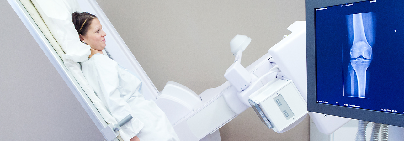
Bone densitometry (or osteodensitometry, or even mineralometry) is a radiological technique that measures bone mineral density. It does so by using a reduced amount of a low-dose X-ray on the bone mass of the site investigated.
It allows for bone calcium deficiencies, such as osteopenia (early stage) or osteoporosis (advanced stage), to be detected. Sites measured are typically the lumbar spine, proximal femurs (hips and femoral necks) and sometimes forearms (radius).
In rare cases, a full-body measurement may also be taken. During the examination, using the same apparatus, lateral spine imaging (vertebral morphometry) is usually carried out in order to look for any vertebral deformations or compression fractures. Furthermore, it is important to be able to assess not just the amount of calcium in the bones (quantitative) but also its distribution (qualitative) by analysing vertebral microarchitecture.
This occurs through a software that, without further acquisition, allows for a vertebral trabecular bone score to be calculated. Depending on all these parameters and a targeted medical questionnaire, the 10-year fracture risk may be determined for all major osteoporotic fractures or hip fractures.
These are essential factors in establishing when and which treatment should be put in place.