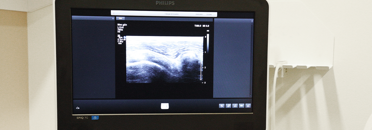
Ultrasound examination in a radiology department.

Ultrasound

This non-radiation and painless medical imaging technique allows a diagnosis to be made or a rapid intervention to be carried out alongside the patient.

Simple, fast and with no contraindications, ultrasound offers many advantages. It can be repeated as often as necessary without any health risks. Ultrasound is very useful in many fields of radiology. This is particularly the case in ENT, to study the salivary glands and thyroid for example, but also in investigating all kinds of organs, whether they are superficial (breasts, musculoskeletal system, blood vessels) or deeper, like organs in the abdomen (liver, gall bladder, pancreas, kidneys, bladder and pelvis).

MRI and ultrasound do not use X-rays

Technically, it is a beam of ultrasound whose frequency is adapted to the organ to be examined. This beam is reflected more or less depending on the anatomical structures it encounters. In turn, this allows for an image of the organ in question to be reconstructed. It is shown directly on the screen of the apparatus for an immediate and dynamic interpretation.

These non-radiation examinations use either the properties of magnetic fields (MRI) or the properties of ultrasound. For the intensities used by these two techniques, no specific effects have been observed in humans.

Examination

At your examination, you will be made comfortable before the radiologist takes over.

At the beginning of the examination, the radiologist applies a gel to the skin above the area to be examined. The gel removes the air between the probe and the skin. This is necessary because the air prevents the transmission of ultrasound.

The examination lasts 15 to 30 minutes depending on the areas being studied.

This technique is also used for targeted biopsies under visual control and as a tracking tool for punctures, drainage and percutaneous treatments.

If preparation is required, do not forget to follow any instructions to ensure that the examination can be carried out correctly and will provide quality results.

Doctors with this specialisation

Clinique de Genolier

Dr. med. Sandrine Schlup-Pidoux

Specialisation
Radiology, Standard radiology, Mammography, MRI, Computed tomography, Ultrasound View more
Clinique de Genolier

Dr. med. Anne-Sophie Knoepfli-Wilson

Specialisation
Radiology, Standard radiology, Mammography, MRI, Computed tomography, Ultrasound View more
Clinique de Genolier

Dr. med. Anne-Lise Hachulla

Specialisation
Radiology, Standard radiology, Mammography, MRI, Computed tomography, Ultrasound View more
Clinique de Genolier

Dr. med. Cathie Kress-Bösch

Specialisation
Radiology, Standard radiology, Mammography, MRI, Computed tomography, Ultrasound View more
Clinique de Genolier

Dr. med. Eric Pesquié

Specialisation
Radiology, Ultrasound, MRI, Computed tomography, Mammography, Standard radiology View more
Clinique de Genolier

Dr. med. Sylvain Eminian

Specialisation
Standard radiology, Radiology, MRI, Computed tomography, Ultrasound, Mammography View more
Clinique de Genolier

Dr. med. Emmanuèle Cerottini

Specialisation
Radiology, Ultrasound, MRI, Mammography, Standard radiology, Interventional radiology, Computed tomography View more
Clinique de Genolier

Dr. med. Yaël Maeder

Specialisation
Radiology, Standard radiology, Mammography, MRI, Computed tomography, Ultrasound View more

Find a doctor

Our specialists have many years of experience. We guarantee a quick, professionally competent clarification and consultation as well as treatment according to the most modern possibilities.