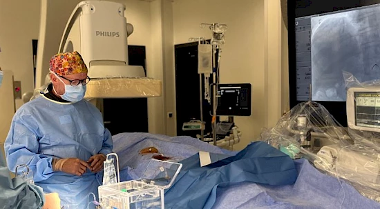
Interventional radiology is a recent medical speciality which relates to both radiology and disciplines such as surgery, oncology and urology in particular. This discipline has the huge advantage of carrying out minimally invasive procedures guided by imaging. It complements surgery for some pathologies, offering new solutions that previously could not be undertaken in radiology or other medical specialities.
From oncology to surgery, here are some of the treatments for which its use is considered invaluable:
IR helps stop bleeding in patients by sealing bleeding vessels from the inside with fewer complications. It also treats haemorrhages, which is particularly important in the postoperative phase. There is no need to operate on the patient again to stop the bleeding.
To destroy tumours, IR uses percutaneous techniques which reach tumours via probes using medical imaging: CT scans, MRI, ultrasound. The aim is to locate the tumour and insert probes into it in order to destroy it. For this purpose, either heat emitted by radio frequencies or microwaves is used. Consequently, the tumour is burned from the inside.
Alternatively, probes that freeze tissues at -80 °C can also be used. The liquid freezes in the cells, which then burst. In this way, the tumours are destroyed without performing an operation or making an incision on the patient. In carefully selected cases, this is as effective as surgery.
Occasionally, IR also offers full cures for tumours that were previously considered incurable. For example, radioactive particles can be injected into a tumour and irradiate it from the inside before eventually destroying it, whatever its type. This is known as radioembolisation, a technique that is well tolerated by patients.
Another very promising technique involves the injection of modified viruses into tumours that will only attack tumour cells. Of course, the virus is modified so that it cannot affect healthy cells.
IR is also used for biliary pathologies, gallstones and bile duct obstructions. Bile ducts are entered to unblock them in the same way as vessels, so that gallstones can be broken up and removed without an operation.
During your examination, you will be accompanied by a medical radiology technologist (MRT). They will provide you with a private area where you can undress according to the requirements of the examination.
If preparation is required, do not forget to follow any instructions to ensure that the examination can be carried out correctly and will provide quality results.
A contrast agent injection may be necessary.
You will be offered a temporary gown.
Please do not hesitate to ask any questions and make any requests. We will strive to fulfil them.
The examination will take 30 minutes on average.